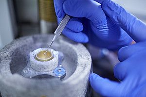
Preparation for cryo-electron microscopy © Charité | Arne Sattler
Charité study in *Science* describes colour-vision pigments in action for the first time
Berlin, June 26, 2026
The human eye can distinguish between hundreds of different colour shades. It achieves this using just three different photopigments—known as colour opsins—located in the cone cells of the retina. Researchers from Charité – Universitätsmedizin Berlin and Nanchang University have now succeeded for the first time in visualizing the molecular structure of all three human colour opsins in their active state. In the journal *Science*, they describe how each of the three opsins reacts to specific light wavelengths, thereby enabling colour vision based on the three primary colours.
The human eye contains around six million cone cells. These are found primarily in the central region of the retina—the fovea—and are responsible not only for colour vision but also for spatial perception and sharp vision in daylight. In dim light or darkness, rod cells located in the outer regions of the retina take over, though they only allow for black-and-white vision. Their photopigment is rhodopsin, the structure of which has been known for 25 years.
Why the structure of colour opsins remained hidden for so long
Cone cells and their corresponding opsins are far less abundant than rhodopsin and are difficult to isolate. “We first had to learn how to produce sufficient quantities of colour opsins in cell cultures, activate them, isolate them, and then examine them using an electron microscope,” explains Dr. Patrick Scheerer. As a doctoral student at Charité’s Institute of Medical Physics and Biophysics in 2008, he played a key role in elucidating the structure of rhodopsin in its active state. “It was a complex process, which is why it took so long for us to finally be able to describe the structures of the colour opsins as well.” Patrick Scheerer now leads the Structural Biology of Cellular Signal Transduction research group at the same institute and is one of the two senior authors of the current study. Like the rhodopsin found in rod cells, all three-colour opsins contain the vitamin A derivative retinal as their light-sensitive component. In each case, it is firmly bound to the opsin protein. When struck by light, it effectively flips at a specific point: the somewhat angled 11-cis-retinal transforms into the elongated all-trans-retinal, thereby altering the shape of the opsin itself and shifting it into an active state. The activated opsin triggers a biochemical signalling cascade that is transmitted via nerve impulses from the eye to the brain for processing—ultimately resulting in colour perception.
Cryo-electron microscopy enables a detailed view
“In this joint project, the three-colour opsins were isolated from cell cultures and—while in their active state, meaning coupled to all-trans-retinal—rapidly cooled to ultra-low temperatures to preserve their active-state structures,” explains Patrick Scheerer, describing the elaborate procedure. Using cryo-electron microscopy, the team was able to generate high-resolution, two-dimensional images at the Ångström scale (sub-nanometre range), making it possible to identify individual amino acids.
From these images, the researchers were able to reconstruct the detailed three-dimensional structure of the opsins. “It turned out that the opsin responding to blue light—that is, short light waves—bears a closer resemblance to the rhodopsin found in rod cells,” reports Patrick Scheerer. “The opsins for green and red light—responding to medium- and long-wavelength stimuli, respectively—differ significantly from this. In particular, the amino acid environment surrounding the retinal varies among the different opsins. This explains their differing sensitivities to specific light wavelengths.” Consequently, colored light stimulates the various cone cells to varying degrees; the combination of these signals and their complex downstream processing gives rise to the perception of color in the brain.
An Important Protein Family for Medicine
All human opsins belong to the protein family known as G-protein-coupled receptors (GPCRs). GPCRs mediate the effects of numerous hormones and neurotransmitters and are involved in a multitude of vital processes, including inflammatory responses, appetite regulation, and growth. Furthermore, they play a central role in sensory perception, such as the detection of odors, tastes, and—of course—light. In fact, rhodopsin was the very first GPCR whose structure was successfully elucidated. In this case, light serves as the stimulus that alters the retinal, thereby activating the GPCR. The more than 800 different GPCRs have long played a pivotal role in both medical applications and the development of new drugs. “Our work is therefore not only scientifically interesting,” says Patrick Scheerer. “There are numerous mutations in opsin genes associated with various eye diseases or visual disorders. Now that we have a better understanding of the structure of opsins, we can classify these mutations more precisely and better understand their effects.”
Also important for the sleep-wake rhythm
For their next project, Patrick Scheerer’s team has turned its attention to melanopsin, which is also found in light-sensitive cells within the retina. These cells transmit signals to the pineal gland and the hypothalamus, thereby playing a crucial role in the sleep-wake cycle. There are also several other opsin genes—likely coding for opsins unrelated to vision. “Our goal is to understand the structural and functional differences between the individual opsins,” says Patrick Scheerer.
*Peng Q et al. Cryogenic electron microscopy structures of human cone visual pigments. Science 2026 Jun 25. doi: 10.1126/science.adz8141
About the study
The study was conducted under the joint leadership of Dr. Patrick Scheerer (Charité) and Prof. Jin Zhang (Nanchang University, China). It also involved scientists from universities in Shenzhen and Ganzhou (China), the Australian National University in Canberra, TU Dortmund University, and the University Alliance Ruhr.
Leave a Reply