Helper protein directs interactive process of ribosome development
The formation process of ribosomes is considered a promising target for potential new antibacterial agents. Researchers at Charité – Universitätsmedizin Berlin have now succeeded in gaining further insights into this multifaceted process. Like in an orchestra, several helper proteins interact with each other during the formation of the ribosome building blocks. Among them is one that guides the entire process like a conductor, the protein ObgE. The team has succeeded in mapping this for the first time. The work has now been published in the journal Molecular Cell*.
Ribosomes are essential components of every cell. They are often referred to as protein factories, because they translate genetic information from the genome into chains of linked amino acids, the proteins. In bacteria, such as the well-known intestinal bacterium Escherichia coli, protein production within the cell – protein biosynthesis – also takes place in this way. If the process fails, the cell dies. Single-celled organisms such as E. coli or other bacteria cannot continue to exist. This fact is to be harnessed in the development of antibiotics. Antibiotic resistance is on the rise, and new multi-resistant germs are emerging and spreading. At the same time, no new classes of antibiotics have been developed for a long time. The aim of novel approaches could be, for example, to intervene in the formation process of ribosomes and block their assembly.
“We happen to be dealing with a viral pandemic at the moment. The next pandemic may well be bacterial in origin, because antibiotic resistance and even multiple resistance are spreading rapidly across species in the bacterial kingdom,” explains Prof. Dr. Christian Spahn, director of the Institute of Medical Physics and Biophysics at Charité and final author of the current study. “The goal of our basic research is therefore to contribute to the development of new antibiotics in the long term.” Together with researchers at the Max Delbrück Center for Molecular Medicine in the Helmholtz Association (MDC) and at the University of Constance, the Charité scientists investigated the question of where exactly in early processes of ribosome development potential targets for new antibacterial and antimicrobial agents can be found.
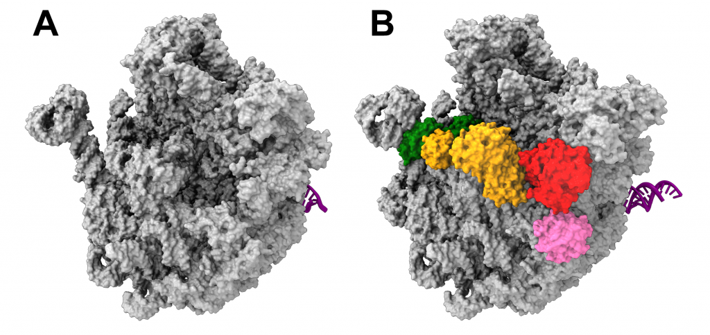
Ribosomes consist of two subunits, one smaller and one larger. The current work of the team led by Dr. Rainer Nikolay, Institute of Medical Physics and Biophysics at Charité, focused on the large ribosomal subunit of the bacterium E. coli and its formation process. As a possible target of novel antibiotics, the scientists wanted to isolate and image precursors of this large subunit – so-called precursors – as natively as possible, i.e. unchanged. For the first time, they have succeeded in taking such a precursor from bacterial cells, in this case E. coli, and imaging the molecular structure at near atomic resolution using cryo-electron microscopy images. “We now better understand at the molecular level, although far from completely, how the large ribosomal subunit forms in a bacterial cell,” said first author Dr. Nikolay.
In order to manipulate the bacterial cell as little as possible for their observations, the research team took a quasi-minimally invasive approach. A key player in the entire process of ribosome development, the protein ObgE, was tagged with a so-called Strep tag. This is done by interfering with the bacterium’s genome, a process known as knock-in. As a result, the bacterium produces only labeled ObgE, which can be visualized by electron microscopy after rapid preparation of the cells. This made it possible for the first time to study the entire complex, because the helper protein ObgE has the precursor of the large ribosomal subunit piggybacking on it, so to speak. With surprising results, as Dr. Nikolay explains: “It turned out that this precursor is covered by numerous helper proteins that interact or communicate with each other. The protein ObgE plays a key role in this by guiding and orchestrating the whole process.” This is precisely where new drugs could come in, blocking the assembly of functional ribosomes and thus preventing bacterial growth.
The team now wants to use similar strategies to gain further insights into the formation process of bacterial ribosomal subunits and to understand the molecular biological processes even better. Work already completed at the Charité and the Max Planck Institute for Molecular Genetics had provided valuable information on the basic structure and different maturation stages of the cellular protein factories. However, these insights were previously based on studies in the test tube, whereas now the formation of the large ribosomal subunit could be reproduced in a living cell. This step is crucial, because in order to identify cellular targets of entirely new pharmaceutical compounds, researchers need to pinpoint differences in the process of ribosome formation in bacteria and in human cells. “We are now a bit closer to doing that,” Dr. Nikolay said. “We’ve been able to reveal both conserved and divergent evolutionary features between prokaryotes – as bacteria are – and eukaryotes – organisms in which the genetic material is present in cell nuclei.” These findings are important for targeting bacteria-specific traits while protecting human cells from unwanted side effects.
*Nikolay R et al. Snapshots of native pre-50S ribosomes reveal a biogenesis factor network and evolutionary specialization. Mol Cell. 2021 Feb 26. doi: 10.1016/j.molcel.2021.02.006.
About the study
The work was supported by the Konstanz Research School Chemical Biology, the Human Frontier Science Program Organization, the German Research Foundation (DFG) Grants FOR1805 and SFB740, the Free University of Berlin and the State of Berlin.
Links:
Institut für Medizinische Physik und Biophysik
Originalpublikation
Pressemitteilung Publikation Mol Cell 2018