Detailed structure of ribosomes in nerve cells elucidated
The finely regulated protein production of the cell takes place at the ribosomes. Which regulators control these processes in certain tissues and in what way? A research team at Charité – Universitätsmedizin Berlin has now investigated this using the detailed structure of the ribosome complex. In this way, the team was able to identify a new regulatory factor for brain development, as now described in the scientific journal Molecular Cell*.
A finely balanced production of proteins – so-called proteostasis – is of great importance, especially for nerve cells. Irregular protein production is therefore a hallmark of many brain diseases. In particular, the early development of the complex cerebral cortex – the neocortex – requires a locally and temporally precisely regulated production of proteins, especially membrane proteins, which play an important role in cell contacts and the linking of nerve cells through synapses. The ribosome is at the centre of this control as a molecular protein factory. Various factors regulate specific protein production in different tissues and at specific times during development. The dynamic interaction of the different factors at the ribosome is largely not understood. However, a research group at the Charité has now succeeded in tracing protein production at the ribosomes in the brain.
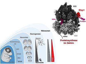
“For the first time, we have been able to visualise the ribosome structure in action in the brain with almost atomic resolution,” says Prof. Dr. Christian Spahn, Director of the Institute for Medical Physics and Biophysics at Charité. “The overall structure of the ribosome has already been elucidated in other tissues or other organisms, but only in this way could we identify Ebp1 as a new central factor that controls ribosome function and the production of certain proteins during brain development.” The regulator protein Ebp1 – the ErbB3 binding protein 1 – contacts the ribosome at its tunnel exit, where a newly emerging protein leaves the ribosome. In this way, the regulator particularly influences the production of membrane proteins that are important for cell contacts and thus maintains neuronal proteostasis.
In a multidisciplinary project, the researchers combined structural biology with neuroscience and combined cryo-electron microscopy (cryo-EM) as a central technique with mass spectrometry, RNA sequencing and genetic methods. With the help of the cryo-EM technique, protein structures – especially large arrangements of several molecules – can be determined at very low temperatures under almost physiological conditions. Neuroscientist Dr. Dr. Matthew L. Kraushar from the Max Planck Institute for Molecular Genetics (MPIMG) in Berlin, first author of the publication and previously a scientist in Prof. Spahn’s laboratory, explains: “This enabled us to look at the molecular architecture of the ribosome in high resolution and as it is present in the nerve cells. In doing so, we were able to capture snapshots of different functional mechanisms of the ribosome.”
“The protein production of the different cell types in the brain is precisely regulated, and small changes can lead to momentous consequences such as neurodegenerative diseases and developmental disorders. With our findings on the role of ribosomes during normal brain development, we will also be able to better understand pathological changes in the brain in the future,” says Prof. Spahn. Next, the researchers want to use large-scale analyses to understand exactly how the ribosome controls the production of the various proteins needed during brain development.
*Kraushar ML et al. Protein synthesis in the developing neocortex at near-atomic resolution reveals Ebp1-mediated neuronal proteostasis at the 60S tunnel exit. Mol Cell. 2020 Dec 22. doi: 10.1016/j.molcel.2020.11.037