Scientists at the Charité – Universitätsmedizin Berlin have shown at the molecular level how light is transformed into information inside cells by means of a protein. In this way, they are broadening our understanding of how bacteria or plants can adapt to different light conditions and control essential processes such as photosynthesis. The findings have been published in the current issue of the journal Nature Communications*.
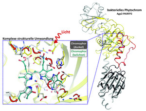
Phytochromes are proteins that are responsible for the conversion of light into information inside cells. These photoreceptors are found in plants, fungi and bacteria and use light to regulate basic physiological processes. Phytochromes contain a light-sensitive molecule, the chromophore. If the chromophore is irradiated with a fixed wavelength of light, it transforms. This is recognized by the surrounding protein and further converted. In the course of activation and deactivation with light, the phytochrome then undergoes a complex structural transformation.
The researchers from the Institute of Medical Physics and Biophysics at the Charité have now contributed to the elucidation of this structural transformation: Using X-ray structure analysis, they determined the 3D structure of a phytochrome in the dark state and compared it with that in the exposed state. To do this, they first converted the protein into a crystalline form and then irradiated it with X-rays. In this way, they were able to calculate the atomic layers of the phytochrome using protein structure analysis. The results show what role individual amino acids play in the activation and deactivation of proteins with light. “Our findings provide basic structural data that contribute to a better understanding of signal transduction from the environment into an organism. Among other things, this knowledge is important for the future use of photoreceptors for various medical applications,” explains Dr. Patrick Scheerer, head of the study.
For example, photoreceptors could be used in oncology for the visualisation of tumour tissue. The basis for this is their ability to absorb and emit red to infrared light. Infrared light can penetrate deeper layers of human tissue, so that even deeper cells can be made visible non-invasively and without side effects with phytochromes. In addition, photoreceptors could be used as light-controlled tools to treat genetic diseases on a molecular level. In order to get closer to these applications, Dr. Scheerer’s team hopes to use the next research projects to better understand the additional fluorescence properties of phytochromes and to investigate further details of the conversion processes of phytochromes.
* Schmidt et al. Structural snapshot of a bacterial phytochrome in its functional intermediate state. Nature Communications. 2018 Nov 21. doi: 10.1038/s41467-018-07392-7